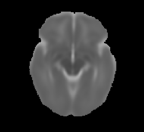
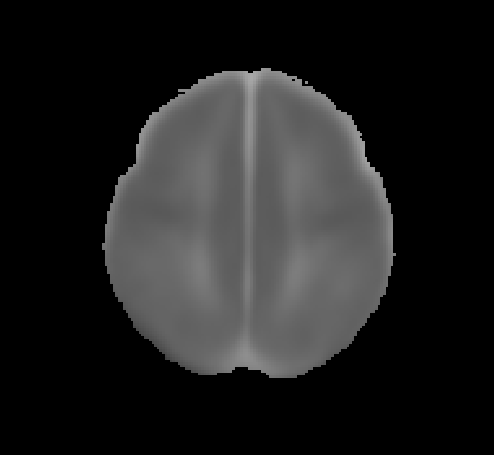
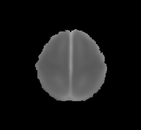
Age-specific map of diffusion metrics in the neonate brain. The data are driven from 565 neonates for DWI/ADC maps, and 162 for DTI metrics. All neonates were neurologically and radiologically normal.
Axial DWIs: TR=6500 ms, TE=90 ms, Flip angle=90, field of view 22x22 cm, slice thickness 4 mm, matrix of 130x130, and b-values of 0 and 1000 s/mm2.
Axial DTIs were obtained using a single-shot, echoplanar imaging sequence: TR=10200 ms, TE=94 ms, Flip angle=90, field of view 18x18 cm, slice thickness 2.5 mm, matrix of 128x128, a single b-0 and 30 b-1000 s/mm2 acquisition
Abbreviations:
FA = Fractional Anisotropy
MD = Mean Diffusivity
ADC = Apparent Diffusion Coefficient
More information
Bobba PS, Weber CF, Mak A, Mozayan A, Malhotra A, Sheth KN, Taylor SN, Vossough A, Grant PE, Scheinost D, Constable RT, Ment LR, Payabvash S. Age-related topographic map of magnetic resonance diffusion metrics in neonatal brains. Hum Brain Mapp. 2022 May 23. doi: 10.1002/hbm.25956. Epub ahead of print. PMID: 35599634.
Version history
Version 2.0 published in February 2022, using a neonate-adapted brain template. Therefore, template images as well as values have changed as compared to Version 1.0
Version 1.0 published June 2021, using standard MNI152 brain templates
Bobba PS, Weber CF, Mak A, Mozayan A, Malhotra A, Sheth KN, Taylor SN, Vossough A, Grant PE, Scheinost D, Constable RT, Ment LR, Payabvash S. Age-related topographic map of magnetic resonance diffusion metrics in neonatal brains. Hum Brain Mapp. 2022 Oct 1;43(14):4326-4334. doi: 10.1002/hbm.25956. Epub 2022 May 23. PMID: 35599634; PMCID: PMC9435001.
Contact website@claraweber.de for technical questions or if you find bugs.
Contributors: Pratheek Bobba, Clara Weber, Adrian Mak, Sam Payabvash
Yale School of Medicine, Department of Radiology and Biomedical Imaging


